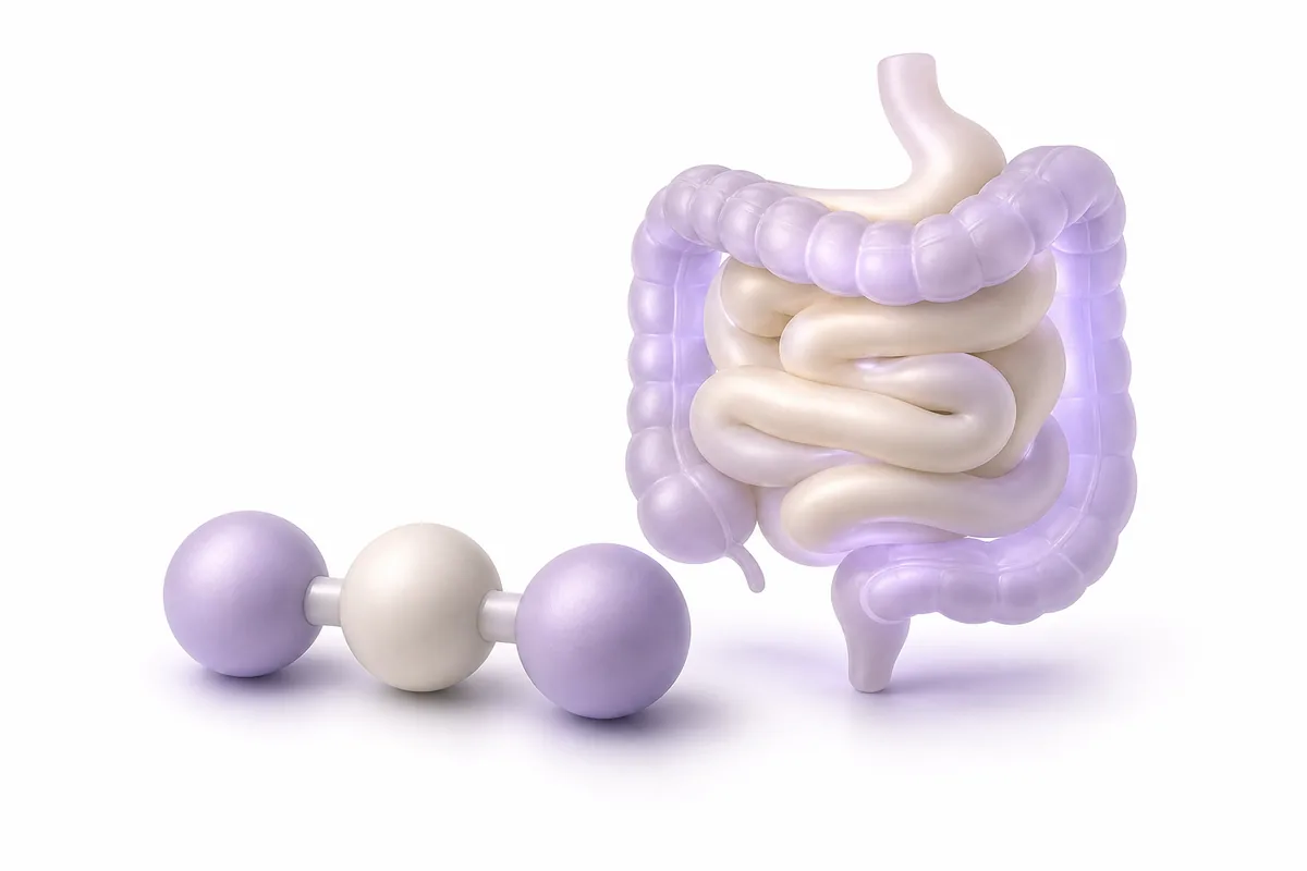
KPV is one of the smallest peptides studied in inflammation research: just three amino acids — lysine, proline, and valine — yet it appears to carry forward much of the anti-inflammatory signaling of its parent hormone, alpha-melanocyte-stimulating hormone (alpha-MSH). This guide summarizes what KPV is, how it is thought to act at the cellular level, and what the preclinical literature — built largely on rodent colitis models and in-vitro cell systems — actually shows. As with all compounds discussed on PeptiMap, everything here is intended strictly for laboratory and educational research purposes, not for human use.
What Is KPV?
Alpha-MSH is a 13-amino-acid peptide hormone best known for its role in skin pigmentation, acting through melanocortin receptors. Alpha-MSH also has a separate anti-inflammatory activity that maps to its C-terminal tripeptide sequence: lysine-proline-valine, positions 11-13 of the parent hormone. This tripeptide is what researchers refer to as KPV.
Because KPV lacks the N-terminal portion of alpha-MSH responsible for melanocortin receptor engagement, it does not appear to produce the pigmentary or appetite-related effects associated with full-length alpha-MSH analogs. Instead, laboratory work has focused almost entirely on its anti-inflammatory and antimicrobial signaling, largely independent of classical melanocortin receptor (MC1R-MC5R) activation — part of what makes it mechanistically distinct from the full alpha-MSH molecule.
Mechanism of Action
Intracellular entry via peptide transporters
Unlike many peptides that act exclusively at the cell surface, KPV’s small size allows it to be taken up directly into cells. In the gut, this uptake is reported to occur substantially through PepT1 (peptide transporter 1), a di/tripeptide transporter normally confined to the small intestine but upregulated in colonic tissue during inflammatory bowel disease. Because PepT1 expression rises specifically at sites of inflammation, its induction may create a route for KPV entry that is more pronounced under inflamed conditions than in healthy colon — a detail researchers have proposed could concentrate the peptide’s activity where it may be more relevant.
NF-κB and IKK inhibition
Once inside the cell, KPV is reported to interfere with the IκB kinase (IKK) complex, a critical upstream regulator of the NF-κB signaling pathway. NF-κB is a transcription factor that, once activated, moves into the nucleus and drives transcription of numerous pro-inflammatory genes, including those encoding TNF-α, IL-6, and IL-1β. By modulating IKK activity, KPV is thought to reduce IκB degradation and, in turn, limit NF-κB nuclear translocation — described in some in-vitro reports as substantially reducing NF-κB activity in stimulated cell systems, though the magnitude varies across models and should not be read as a fixed, universal figure.
MAPK pathway involvement
Alongside NF-κB, some laboratory reports describe KPV as also modulating mitogen-activated protein kinase (MAPK) signaling, a parallel pathway implicated in cytokine production. Together, these two intracellular actions are proposed as the basis for the broad anti-inflammatory profile observed for KPV in cell and animal studies.
Receptor-independent and receptor-mediated components
Earlier work using immobilized alpha-MSH C-terminal peptide constructs suggested a more receptor-mediated component to some of these effects in certain cell systems, indicating that KPV’s full mechanism likely involves more than one pathway and is still being worked out (Brzoska et al., 2010). Overall, the mechanistic picture is best described as intracellular and multi-pathway, rather than fully resolved.
Research Background and Key Findings
KPV has been examined across several preclinical contexts:
- Colitis models. In rodent models of colitis induced by dextran sodium sulfate (DSS) and TNBS (2,4,6-trinitrobenzenesulfonic acid), oral administration of KPV has been reported to reduce measures of disease severity, including inflammatory markers and mucosal damage scores (Dalmasso et al., 2008).
- Targeted colonic delivery. Follow-on work exploring colon-targeted delivery systems for KPV has reported reductions in TNF-α expression alongside indications of accelerated mucosal healing in ulcerative-colitis-type mouse models.
- Skin and dermal inflammation. Older work on alpha-MSH C-terminal fragments — the structural family KPV belongs to — found reduced contact hypersensitivity responses in dermal models, a line of research relevant to how suppressing local inflammation could, in principle, support wound-related repair processes (Brzoska et al., 2010).
- Transdermal delivery research. Because KPV is a small, charged tripeptide, delivery-focused pharmaceutical science has also studied how it might be moved across skin barriers using methods such as iontophoresis and microporation, independent of any therapeutic claim (Pawar et al., 2017).
- Broader antimicrobial and anti-inflammatory profile. Review literature on alpha-MSH-related peptides situates KPV within a wider family of compounds studied for combined anti-inflammatory and antimicrobial activity across inflammatory disease models (Singh & Aggarwal, 2014).
Across all of this work, a consistent caveat applies: no large controlled human clinical trials of KPV have been published. The evidence base remains preclinical — rodent colitis models, in-vitro cell assays, and skin-permeation studies — and findings in these systems should not be interpreted as proof of any effect in humans. For related preclinical anti-inflammatory and repair-oriented peptide research, see our overview of BPC-157, another peptide frequently discussed in gut- and tissue-focused research contexts.
Forms, Reconstitution & Handling
In research settings, KPV is typically supplied as a lyophilized (freeze-dried) powder in sealed vials, such as the KPV 5mg format. As with other lyophilized peptides, it must be reconstituted with an appropriate diluent — commonly bacteriostatic water — before it can be used in laboratory work.
General handling considerations that apply broadly to small peptides like KPV include:
- Allowing the vial to reach room temperature before reconstitution to reduce mechanical stress on the lyophilized cake.
- Adding diluent slowly along the interior wall of the vial rather than directly onto the powder, and swirling gently rather than shaking, to minimize the risk of degradation.
- Labeling vials clearly with reconstitution date, diluent volume, and resulting concentration to support reproducible record-keeping.
- Working in a clean, low-light environment, since peptide stability can be sensitive to light and temperature fluctuations.
For a general walkthrough of reconstitution calculations and technique, see our peptide reconstitution guide.
Research Considerations
Because KPV is under active study across multiple delivery routes — oral, topical, and injectable, in various preclinical designs — researchers approaching this peptide should keep several general points in mind:
- Route matters mechanistically. Since PepT1-mediated uptake in the gut is central to several proposed mechanisms, oral and topical/enteric delivery models are handled very differently from systemic (injectable) approaches in the literature, and findings from one route should not automatically be assumed to generalize to another.
- Model heterogeneity. DSS-colitis, TNBS-colitis, and dermal hypersensitivity models each represent distinct disease processes; results in one model do not necessarily predict results in another.
- Formulation and stability in solution. As a very small, unprotected tripeptide, KPV may be more susceptible to enzymatic degradation than larger, more structurally stabilized peptides, a variable that research protocols typically need to account for.
- Reporting rigor. As with any actively studied compound, researchers should evaluate primary literature directly rather than relying on secondary summaries, and should be cautious of sweeping claims not tied to a specific study design.
This section is general research context, not a protocol or instruction for use.
Storage & Stability
- Lyophilized form: Store frozen (around −20°C), protected from light and moisture, where it is generally reported to remain stable for extended periods.
- Reconstituted form: Refrigerate (roughly 2-8°C) and avoid extended storage; many labs default to using reconstituted small peptides within a few weeks, consistent with general practice for compounds of this size.
- Avoid freeze-thaw cycling. Repeated temperature swings can accelerate degradation of small peptides in solution.
- Protect from light. UV and prolonged ambient light exposure are commonly cited risk factors for peptide degradation during storage.
See our peptide storage guide for a broader discussion of handling practices across peptide classes.
Safety, Legality & Research Disclaimers
KPV is a research chemical, not an approved drug. It has no marketing authorization as a medicine in the EU, the United States, or other major jurisdictions, and its legal and regulatory status can vary by country and may change over time. Researchers are responsible for confirming current regulations in their own jurisdiction and for complying with their institution’s ethics, biosafety, and procurement requirements.
Available preclinical safety data on KPV is limited primarily to short-term animal and cell-based studies; comprehensive long-term human safety information does not exist because controlled human trials have not been conducted. Nothing in this article constitutes medical advice, a recommendation for human use, or an endorsement of self-administration. All material is intended strictly for qualified laboratory research and educational purposes.
Frequently Asked Questions
Is KPV approved for human use? No. KPV is an investigational research compound with no regulatory approval as a medicine. It is intended for laboratory research only.
How is KPV different from alpha-MSH? KPV is only the C-terminal three-amino-acid fragment of the 13-amino-acid alpha-MSH hormone. It appears to retain much of alpha-MSH’s anti-inflammatory signaling while lacking the receptor-binding region responsible for the pigmentation and appetite effects associated with full-length alpha-MSH.
What does “preclinical” mean in the context of KPV research? It means the existing evidence for KPV’s anti-inflammatory activity comes from rodent models (such as DSS- and TNBS-induced colitis) and in-vitro cell studies, not from large controlled human clinical trials. Preclinical results are informative but cannot be assumed to translate directly to people.
Why is PepT1 relevant to KPV research? PepT1 is a di/tripeptide transporter that is upregulated in the colon during inflammatory bowel disease. Because KPV is small enough to be a substrate for this transporter, PepT1-mediated uptake has been proposed as a route by which KPV may enter inflamed intestinal cells more readily than healthy tissue — a detail of mechanistic interest to gut-inflammation researchers.
Is KPV studied alongside other research peptides? Yes — reviewers and researchers frequently discuss KPV in the same anti-inflammatory and tissue-repair research context as other preclinical peptides, such as BPC-157, even though the two have distinct proposed mechanisms.
Where can I find product-specific details for research use? General vial information for laboratory use is available on the KPV 5mg page. This is not dosing guidance for human use.
References
- Dalmasso G, Charrier-Hisamuddin L, Nguyen HTT, Yan Y, Sitaraman S, Merlin D. “PepT1-Mediated Tripeptide KPV Uptake Reduces Intestinal Inflammation.” Gastroenterology. 2008.
- Brzoska T, Böhm M, Lügering A, Loser K, Luger TA. “Terminal Signal: Anti-Inflammatory Effects of α-Melanocyte-Stimulating Hormone Related Peptides Beyond the Pharmacophore.” Advances in Experimental Medicine and Biology. 2010.
- Pawar K, Kolli CSR, Rangari VK, Babu RJ. “Transdermal Iontophoretic Delivery of Lysine-Proline-Valine (KPV) Peptide Across Microporated Human Skin.” Journal of Pharmaceutical Sciences. 2017;106(7):1814-1820.
- Singh S, Aggarwal A. “Alpha-Melanocyte Stimulating Hormone: An Emerging Anti-Inflammatory Antimicrobial Peptide.” BioMed Research International. 2014.