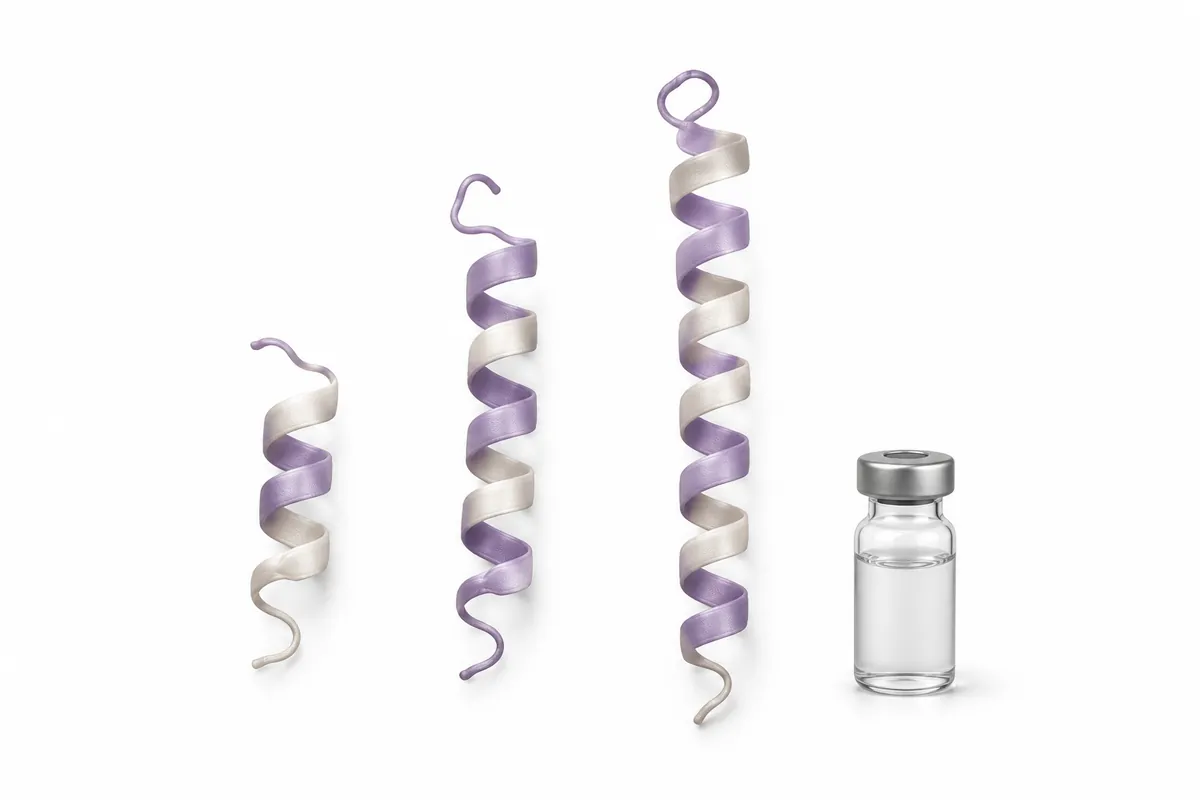
TL;DR: IGF-1 LR3, IGF-1 DES, and PEG-MGF are three IGF-1-family research peptides with very different pharmacokinetics. LR3 is a long-acting, systemic IGF-1 analogue; DES is a truncated, shorter-acting form that is more potent locally; PEG-MGF is a PEGylated splice variant aimed at local satellite-cell activation after mechanical damage. All three are evidence-poor, with most claims extrapolated from IGF-1 biology rather than dedicated human trials.
The core difference: same family, three PK strategies
All three compounds trace back to insulin-like growth factor 1 (IGF-1), a hormone central to muscle growth, tissue repair, and cellular proliferation. What separates them is not the downstream biology — they all ultimately touch IGF-1 signalling — but where and for how long that signal is delivered.
IGF-1 LR3 (Long R3 IGF-1) is a modified analogue with an arginine substitution at position 3 and a 13-amino-acid extension at the N-terminus. Those changes make it resistant to binding by IGF-binding proteins (IGFBPs), which normally sequester circulating IGF-1. Escaping IGFBP capture means more free peptide, a much longer half-life, and a systemic reach.
IGF-1 DES (Des1-3 IGF-1) goes the other way: it is a truncated form missing the first three N-terminal amino acids. That deletion sharply lowers its affinity for IGFBPs too, but because it is not stabilised, it clears quickly. Its signature is high local potency over a short window.
PEG-MGF is different in kind. MGF (mechano growth factor, also called IGF-1Ec) is a splice variant of the IGF-1 gene expressed by muscle in response to mechanical loading or damage. It is thought to act locally to activate satellite cells and kick off repair. Native MGF has a half-life measured in minutes; PEGylation — attaching a polyethylene glycol chain — extends that window so the molecule persists long enough to study.
You can review individual reference profiles where catalogued, and compare against related repair-focused compounds such as TB-500 and the myostatin-adjacent Follistatin 344.
Mechanism: one receptor, different delivery
IGF-1 LR3 and IGF-1 DES both act as agonists at the IGF-1R (IGF-1 receptor), a tyrosine kinase that, once engaged, drives the PI3K/Akt/mTOR cascade associated with protein synthesis and cell survival, plus the Ras/MAPK arm associated with proliferation. In muscle research, that signalling is linked to satellite-cell activation and mitogenesis — the machinery behind hypertrophy and repair.
The practical distinction is IGFBP handling. Roughly 98% of endogenous IGF-1 circulates bound to IGFBPs, which act as a reservoir and brake. Both LR3 and DES are engineered to slip that brake, but LR3’s N-terminal extension also protects it from clearance, so it keeps signalling systemically for a long time. DES clears fast, concentrating its effect near the injection site before it degrades.
PEG-MGF’s mechanism is less about classic IGF-1R agonism and more about satellite-cell dynamics. MGF is thought to signal through a pathway distinct from mature IGF-1 — its unique E-domain peptide appears to stimulate satellite-cell proliferation and delay their differentiation, expanding the pool of cells available for repair after mechanical damage. Mature IGF-1 (and analogues like LR3) is then associated with pushing those cells toward differentiation and fusion. In that framing, MGF and the mature IGF-1 forms are studied as sequential, complementary steps rather than substitutes.
Half-life and duration: the sharpest contrast
Half-life is where these three diverge most, and it drives every downstream difference in how they are studied.
Approximate research figures, not clinical values. IGF-1 LR3 persists for a day-plus; DES and native MGF clear in minutes, with PEGylation extending MGF.
IGF-1 LR3 is the long-acting member. Its IGFBP resistance and clearance-resistant structure give it an often-cited half-life on the order of 20–30 hours, which is why it is discussed in terms of systemic, sustained IGF-1R exposure and infrequent administration.
IGF-1 DES is the short-acting counterpart, with a half-life commonly cited around 20–30 minutes. Its research profile is built around brief, high-intensity local signalling — a spike near the site rather than a systemic bath.
PEG-MGF sits in between by design. Native MGF’s E-domain is degraded within minutes, which would make it almost impossible to work with; the PEG chain shields it and stretches the functional window into hours, long enough for the local satellite-cell effect to be studied without minute-by-minute redosing.
| Attribute | IGF-1 LR3 | IGF-1 DES | PEG-MGF |
|---|---|---|---|
| Full name | Long R3 IGF-1 | Des1-3 IGF-1 | PEGylated Mechano Growth Factor |
| Structure | IGF-1 + Arg3 sub + 13-aa N-term extension | IGF-1 minus first 3 N-term residues | IGF-1Ec splice variant + PEG chain |
| Primary receptor / target | IGF-1R (systemic) | IGF-1R (local) | Satellite cells (local repair) |
| Signalling associated with | PI3K/Akt/mTOR, Ras/MAPK | PI3K/Akt/mTOR (local) | Satellite-cell proliferation |
| IGFBP binding | Very low (resistant) | Very low (resistant) | Not the primary axis |
| Approx. half-life | ~20-30 hours | ~20-30 minutes | Hours (PEG-extended) |
| Action mode | Systemic, sustained | Local, brief, potent | Local, mechanical-damage repair |
Local vs systemic: what each is studied for
The half-life and structural choices map cleanly onto research use-cases.
IGF-1 LR3 — systemic reach. Because it circulates freely and persists, LR3 is the form associated with whole-body IGF-1R signalling. Research discussion centres on systemic hypertrophy models and sustained mTOR activation. The flip side of systemic potency is that IGF-1R is expressed almost everywhere, so continuous whole-body activation is exactly the property that makes its long-term profile hard to characterise.
IGF-1 DES — local potency. DES is studied where a strong, brief, site-concentrated pulse is the point. Its low IGFBP affinity and short half-life mean it is often described as more potent locally than native IGF-1 on a per-molecule basis, but without the systemic persistence of LR3. In practice, that makes it the form researchers reach for when they want to probe a localised response.
PEG-MGF — repair after mechanical damage. PEG-MGF’s research niche is the window right after mechanical loading or injury, when native MGF would naturally spike. The hypothesis under study is that supplying a longer-lasting MGF analogue expands satellite-cell recruitment during that repair window. It is a local-repair story, not a systemic-growth one.
Evidence quality: be honest — it’s thin
This is the part that matters most, and it applies to all three roughly equally.
A few specifics worth stating plainly:
- IGF-1 LR3 has a solid mechanistic and industrial pedigree — it is well characterised as a potent IGF-1R agonist in vitro and is widely used to sustain cell cultures. Its use as a systemic muscle-growth agent in humans is not supported by dedicated trials; that application is extrapolation.
- IGF-1 DES is real and well-described biochemically, including as a naturally occurring IGF-1 variant in some tissues. Its “more potent locally” reputation rests largely on receptor-binding and in-vitro data, not human outcome studies.
- PEG-MGF rests on genuine MGF splice-variant biology (the IGF-1Ec transcript is well documented), but the PEGylated research compound itself has little dedicated human evidence. Much of what is claimed comes from rodent and cell-based satellite-cell work.
None of the three is an approved medicine. Treating any of them as an established, characterised intervention overstates what the literature actually shows. The honest summary is that the IGF-1 axis is well studied; these specific analogues as performance or repair agents are not.
How to think about choosing between them
Because the evidence is thin across the board, “which is best” is not a question the literature can answer. What it can offer is a clean mechanistic fit:
- Studying sustained, systemic IGF-1R signalling points to IGF-1 LR3 — the only long-acting, systemically distributed option here.
- Probing a brief, potent, localised IGF-1 response points to IGF-1 DES, whose short half-life keeps the effect near the site.
- Modelling satellite-cell activation after mechanical damage points to PEG-MGF, which targets the repair window rather than systemic growth.
Some study designs examine PEG-MGF alongside a mature IGF-1 form on the reasoning that they occupy different steps of the repair sequence. For broader context on how researchers combine repair-oriented peptides, see our common peptide stacks overview and the CJC-1295 + ipamorelin blend discussion, which covers the growth-hormone axis that sits upstream of endogenous IGF-1.
Frequently Asked Questions
What is the main difference between IGF-1 LR3 and IGF-1 DES?
Both are IGFBP-resistant IGF-1R agonists, but their pharmacokinetics are opposite. IGF-1 LR3 has an N-terminal extension that makes it long-acting and systemic, with a half-life on the order of 20–30 hours. IGF-1 DES is truncated and short-acting, clearing in roughly 20–30 minutes, which concentrates its effect locally rather than systemically.
Is PEG-MGF the same as IGF-1?
No. MGF (mechano growth factor, or IGF-1Ec) is a splice variant of the IGF-1 gene, not mature IGF-1. It carries a unique E-domain associated with local satellite-cell activation after mechanical damage, and it is studied as an early repair signal rather than a systemic growth driver. PEGylation is added to extend its otherwise very short half-life.
Why is IGF-1 DES considered more potent locally?
DES has very low affinity for IGF-binding proteins, so more of it is available to engage IGF-1R near the injection site, and receptor-binding data suggest a strong per-molecule effect. Combined with its short half-life, that produces a brief, intense local signal rather than the sustained systemic exposure seen with LR3. Note this “more potent” framing rests mainly on in-vitro data.
How strong is the human evidence for these three peptides?
Weak. Most claims are extrapolated from general IGF-1 biology, cell culture, and animal models rather than controlled human trials of these specific analogues. IGF-1 LR3 in particular was developed largely as a cell-culture supplement. None of the three is an approved medicine, and their muscle- or repair-related applications in humans are inference, not demonstrated fact.
Are IGF-1 LR3, DES, and PEG-MGF interchangeable?
No. They differ in structure, half-life, and where they act — LR3 is long-acting and systemic, DES is short-acting and local, and PEG-MGF targets local satellite-cell repair through a distinct splice-variant pathway. They answer different research questions and should be treated as separate reference materials, not substitutes.
References
- Francis GL, Ross M, Ballard FJ, et al. Novel recombinant fusion protein analogues of insulin-like growth factor (IGF)-I indicate the relative importance of IGF-binding protein and receptor binding for enhanced biological potency. Journal of Molecular Endocrinology. 1992;8(3):213-223.
- Ballard FJ, Wallace JC, Francis GL, et al. Des(1-3)IGF-I: a truncated form of insulin-like growth factor-I. The International Journal of Biochemistry & Cell Biology. 1996;28(10):1085-1087.
- Yang S, Alnaqeeb M, Simpson H, Goldspink G. Cloning and characterization of an IGF-1 isoform expressed in skeletal muscle subjected to stretch. Journal of Muscle Research and Cell Motility. 1996;17(4):487-495.
- Hameed M, Orrell RW, Cobbold M, Goldspink G, Harridge SD. Expression of IGF-I splice variants in young and old human skeletal muscle after high resistance exercise. The Journal of Physiology. 2003;547(1):247-254.
- Philippou A, Maridaki M, Halapas A, Koutsilieris M. The role of the insulin-like growth factor 1 (IGF-1) in skeletal muscle physiology. In Vivo. 2007;21(1):45-54.
- Tomas FM, Lemmey AB, Read LC, Ballard FJ. Superior potency of infused IGF-I analogues which bind poorly to IGF-binding proteins. Journal of Endocrinology. 1996;150(1):77-84.
This article is provided for scientific and educational reference only. All compounds discussed are intended strictly for in-vitro laboratory research use. Nothing here is medical advice, and no statement should be read as a therapeutic claim, an endorsement, or a human dosing recommendation. IGF-1 LR3, IGF-1 DES, and PEG-MGF are not approved for human consumption.