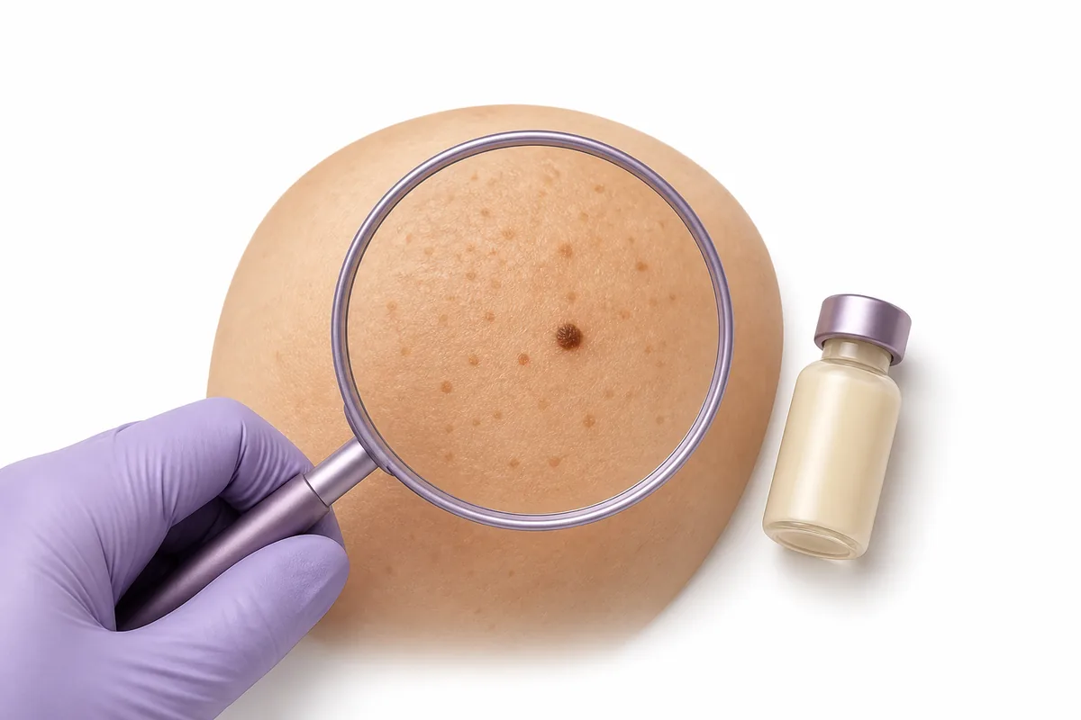
TL;DR: Melanotan II darkens existing pigment because it activates MC1R on melanocytes, pushing them to make more eumelanin — the same pathway responsible for the tan. That pathway doesn’t distinguish ordinary skin from a mole or freckle, so pigmented lesions darken along with everything else, and faint ones can become visible enough to look “new.” Whether MT-II also drives genuinely new lesion formation or atypical change is a separate question, and the evidence there is case-report tier only — a handful of published clinical cases, not controlled trials, confounded by the fact that people using MT-II are usually also chasing UV exposure.
The question everyone asks
A few days into a Melanotan II course, a familiar post shows up in research-peptide forums: people notice what looks like more freckles and moles than before, almost as if new ones are forming, and ask whether this is just existing skin quirks darkening or something worth watching. It’s one of the most repeated questions in that community, and it deserves a real answer.
Short version: darkening of pigment you already had is the expected, mechanistically predictable effect. Whether MT-II also creates genuinely new lesions, or drives atypical change in existing ones, is a harder question — one where real evidence exists but it’s thin. Here’s both, and where the line between them sits.
The mechanism: MC1R, eumelanin, and darkening pigment
Melanotan II is a non-selective melanocortin receptor agonist — it activates MC1R, MC3R, MC4R, and MC5R rather than one receptor alone. Our companion piece on what Melanotan II is covers the full receptor picture, including the MC4R-driven appetite and arousal effects along for the ride. For moles and freckles, MC1R is the one that matters.
MC1R sits on melanocytes, the pigment-producing cells across skin. Activating it triggers a cAMP-mediated cascade that upregulates MITF, which raises tyrosinase activity and shifts production toward eumelanin — the darker, brown-black pigment. That’s the tanning mechanism in miniature, and it’s well characterized pharmacologically.
The part that matters here: melanocytes inside a mole or freckle are still melanocytes. MC1R activation doesn’t check whether a patch of skin is a nevus, an ephelis, or ordinary surrounding skin before switching on eumelanin production. A mole that was already there darkens for the same reason the rest of the skin does. A freckle too faint to notice before can cross the threshold into visible. That plausibly explains a meaningful share of “new mole” reports: not a lesion appearing from nothing, but an existing, previously-inconspicuous one becoming visible.
Where the mechanism runs out of clean answers: whether sustained melanocortin activation can also drive melanocyte proliferation — actual new nevus formation, or a stable nevus transforming into something atypical — is genuinely open. A 2025 pharmacology review in the Journal of the European Academy of Dermatology and Venereology notes MC1R signaling normally participates in controlling aberrant cell growth, but is explicit that MC1R activation does not prevent melanoma, particularly in people with existing risk factors, and that regular skin exams remain important for anyone in an elevated-risk category. Pharmacological activation outside normal physiological control isn’t simply equivalent to sun-driven tanning.
Melanotan I vs. Melanotan II: the selectivity that changes everything
Melanotan I — developed into the approved drug afamelanotide, marketed as Scenesse — is MC1R-selective: it activates the pigmentation receptor at doses well below what would meaningfully engage MC3R/MC4R/MC5R. That selectivity is why afamelanotide became an approved drug. The FDA approved it in 2019 and the EMA in 2014, specifically to prevent phototoxic reactions in adults with erythropoietic protoporphyria (EPP), a rare severe light-sensitivity condition. It’s delivered as a controlled-release subcutaneous implant under clinical supervision, not self-injected.
Melanotan II never went through that process and has no approved medical indication anywhere. Its non-selectivity is the source of both its broader side-effect profile (nausea, flushing, spontaneous arousal, appetite changes) and its lack of any built-in ceiling on how it engages pigment-producing cells throughout the body — moles included.
What the case-report literature actually documents
Precision on evidence tier matters here. There is no randomized trial data on MT-II and mole change in humans — nobody has run a controlled study exposing volunteers to MT-II and tracking nevus outcomes against placebo. What exists instead is a small cluster of individual case reports in dermatology journals, each describing one or a few patients who self-administered Melanotan and then presented with some form of pigmented-lesion change.
- 1
2009 — British Journal of Dermatology
Cousen, Colver & Helbling describe eruptive melanocytic naevi following melanotan injection, reporting rapid pigmentation and new nevi, and reference an earlier case of rapidly developing melanoma in a melanotan user.
- 2
2011 — British Journal of Dermatology
Paurobally et al. report "Melanotan-associated melanoma": a 42-year-old woman developed an enlarging, darkening abdominal mole over three months of MT-II use; excision found a thin (0.30mm Breslow) melanoma. The authors note a temporal association without establishing causation.
- 3
2012 — Actas Dermo-Sifiliográficas
Hueso-Gabriel et al. describe a 25-year-old man who developed more than 100 melanocytic nevi within weeks of a four-week MT-II course; ten excised lesions were dysplastic, three severely so.
- 4
Ongoing
DermNet NZ's clinical summary on Melanotan II lists deepening of existing moles, new moles, and atypical melanocytic naevi among reported concerns, alongside a documented FAMMM-syndrome case covered in our companion Melanotan II article.
Reading across these, a pattern holds: darkening and enlargement of existing lesions appears in essentially every case, and a subset also describe new lesions and, less often, dysplastic or malignant histopathology. What none of these reports can do, by design, is establish a causal rate. Case reports are the weakest tier of clinical evidence for causation — they describe one identifiable patient, not an average. And nearly every published case involves someone using Melanotan specifically to tan, meaning concurrent UV exposure is baked into the record — the biggest confound here, since sun exposure is an independently established melanoma risk factor on its own.
So: darkening of existing pigment is expected pharmacology, well explained by MC1R biology. New lesions and the rarer dysplastic or malignant reports are real observations in the literature, but sit at case-report tier, confounded by UV exposure, and a causal link to melanoma is not established.
The ABCDE reference, for context
Dermatology uses a standard framework for describing what a changing or atypical mole can look like. It’s included here as background, not a self-diagnosis checklist:
Anyone with a personal or family history of atypical nevi, dysplastic nevus syndrome, or melanoma sits in a different risk category, and the FAMMM-syndrome case in our Melanotan II overview shows why that history matters more here — MC1R activation reaching a genetically primed population of atypical melanocytes is a different scenario than reaching an unremarkable one.
Where this leaves the picture
MT-II’s effect on existing pigmented lesions isn’t a mysterious side effect — it follows directly from the MC1R → eumelanin pathway that produces the tan, applied indiscriminately to every melanocyte it reaches, moles and freckles included. That’s well-supported pharmacology. Whether it also drives new melanocytic proliferation, and whether that occasionally intersects with dysplastic or malignant transformation, is where evidence thins to a handful of case reports — real and worth knowing, but not the kind of evidence that supports a firm incidence estimate or settled causal story. For sourcing and research-material quality more broadly, our peptide quality and safety guide and vendor-vetting guide are useful background, and peptide dosing 101 covers how concentration and unit math work across any compound.
Frequently asked questions
Does Melanotan II actually create new moles, or just darken old ones?
Both are reported, but not equally well explained. Darkening of existing pigment follows directly from MC1R-driven eumelanin production and is well-supported mechanistically. Whether MT-II also drives genuinely new melanocyte proliferation is less certain — a previously faint mole becoming obviously dark can look identical to a “new” mole appearing, and most people don’t have a before photo to tell the two apart.
Is the mole-darkening reported with Melanotan II dangerous?
Darkening alone, following the expected MC1R mechanism, isn’t automatically a danger sign — it’s the same pathway responsible for tanning. But since a small number of case reports describe dysplastic or malignant change in pigmented lesions during Melanotan use, any lesion changing in asymmetry, border, colour, diameter, or otherwise evolving is worth having examined, using ABCDE as a reference point rather than a self-diagnosis tool.
What’s the difference between Melanotan I and Melanotan II for skin pigmentation?
Melanotan I, developed into the approved drug afamelanotide (Scenesse), is MC1R-selective, activating the pigmentation receptor without meaningfully engaging other melanocortin receptors at typical doses. Melanotan II is non-selective, activating MC1R alongside MC3R, MC4R, and MC5R, which is why it carries a broader side-effect profile and no approved medical indication.
Has a causal link between Melanotan II and melanoma been established?
No. The published literature consists of individual case reports — the weakest tier of clinical evidence for causation — and nearly all involve concurrent UV/tanning exposure, an independent, well-established melanoma risk factor on its own. A temporal association has been reported in a small number of cases; a controlled causal link has not.
Who should be more cautious based on the case-report literature?
People with a personal or family history of atypical nevi, dysplastic nevus syndrome, or melanoma appear in the case-report literature at what looks like disproportionate representation, including a documented case involving FAMMM syndrome. That history changes the risk calculus more than it does for someone without it.
Should I get a mole checked if it changes during a Melanotan course?
That’s a question for a clinician examining the actual lesion, not something answerable in the abstract. ABCDE is the standard reference dermatologists use to describe a changing or atypical lesion, provided here as background context rather than a substitute for an in-person exam.
References
- Cousen, P., Colver, G., & Helbling, I. (2009). Eruptive melanocytic naevi following melanotan injection. British Journal of Dermatology, 161(3), 707–708.
- Paurobally, D., Jason, F., Dezfoulian, B., & Nikkels, A. F. (2011). Melanotan-associated melanoma. British Journal of Dermatology, 164(6), 1403–1405.
- Hueso-Gabriel, L., Mahiques Santos, L., Terrádez Mas, L., & Santonja López, C. (2012). Eruptive Dysplastic Nevi Following Melanotan Use. Actas Dermo-Sifiliográficas, 103(4), 329–331.
- Böhm, M., Robert, C., Malhotra, S., Clément, K., & Farooqi, S. (2025). An overview of benefits and risks of chronic melanocortin-1 receptor activation. Journal of the European Academy of Dermatology and Venereology, 39(1), 39–51.
- DermNet NZ. Melanotan II. Retrieved from https://dermnetnz.org/topics/melanotan-ii
- U.S. Food and Drug Administration. Afamelanotide (Scenesse) approval, 2019, for prevention of phototoxicity in adults with erythropoietic protoporphyria.
Disclaimer: This information is for educational and research purposes only. Peptides are research chemicals not intended for human consumption. Nothing in this article is medical advice; any change in a mole or pigmented lesion should be evaluated by a qualified clinician.